

Ear Keloid
Earlobe Reduction & Repair
Blepharoplasty
Mole, Cyst & Lipoma Removal
Liquid Rhinoplasty
Neck Contouring
FACE GALLERY
EAR KELOID
Ear Keloid Before & After Photos
Ear keloids can vary considerably in size, shape and location. They may develop on the earlobe or cartilage portion of the ear, often following an ear piercing. These ear keloid before and after photos show examples of patients treated by Dr. Adelyn Ho. Treatment is individualized according to the keloid, surrounding ear tissue, previous treatment and risk of recurrence. Learn more about ear keloid removal in Vancouver, including surgery, steroid injections, pressure therapy, recurrence and recovery.
BEFORE
AFTER
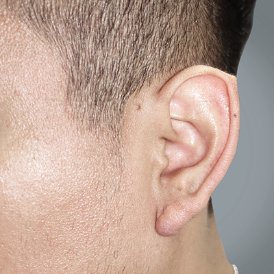
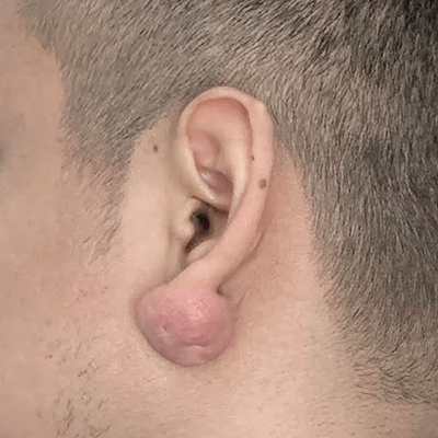
Patient No. 1 — Ear Keloid Before & After: Large Earlobe Keloid After Piercing
This patient presented with a large earlobe keloid after an ear piercing. Because of its size, treatment required surgical excision with careful closure to preserve the shape of the earlobe. Postoperative care included multiple steroid injections and pressure earring therapy. The after photo is shown at 3 months.
BEFORE
AFTER
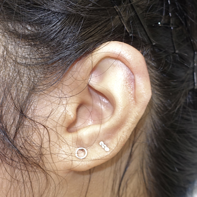
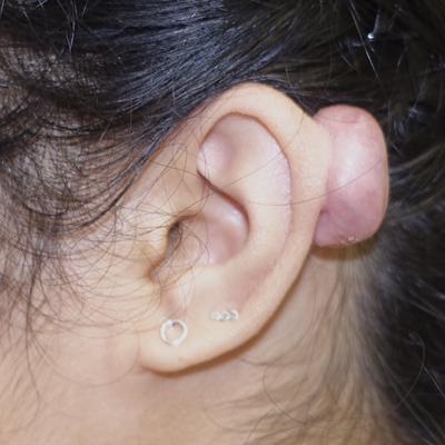
Patient No. 2 — Ear Keloid Before & After: Cartilage Keloid After Piercing
This patient developed a large keloid on the cartilage portion of the ear after a piercing. The keloid projected behind the upper ear and required treatment that considered both removal of the scar and preservation of the surrounding ear contour. The after image shows restoration of the upper ear shape following treatment.
BEFORE
AFTER
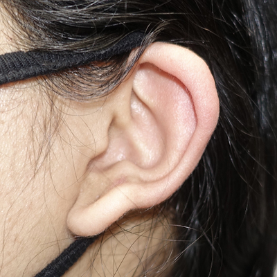
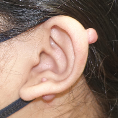
Patient No. 3 — Ear Keloid Before & After: Multiple Small Ear Keloids
This patient had several smaller keloids involving different areas of the ear, including the upper helical rim and lower ear. The after photograph shows a smoother ear contour following treatment of the raised scars.
BEFORE
AFTER
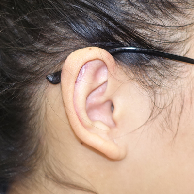
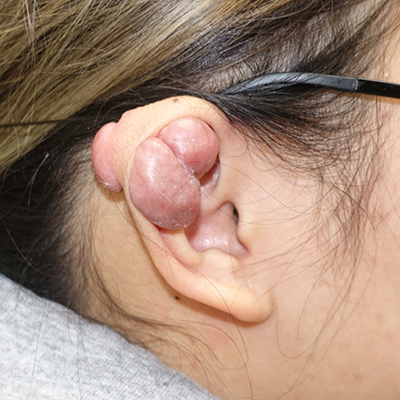
Patient No. 4 — Ear Keloid Before & After: Very Large Cartilage Keloid
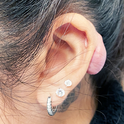
This patient presented with a very large keloid involving the upper cartilage and helical rim of the ear. Large cartilage keloids require careful planning because treatment must address the scar while preserving the shape and supporting structure of the ear. Steroid treatments and pressure earring therapy was followed compliantly.
BEFORE
AFTER
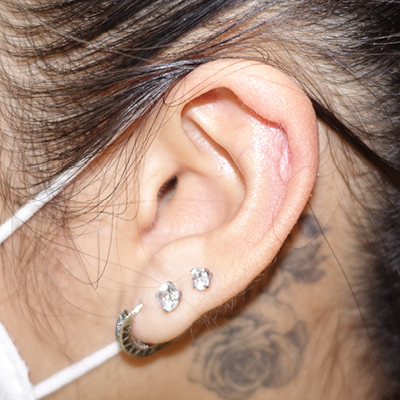
Patient No. 5 — Ear Keloid Before & After: Posterior Cartilage Keloid
This patient underwent ear keloid removal for a keloid that developed after piercing..The before and after photographs demonstrate the change in the contour of the helical rim following treatment of the posterior keloid with surgery and steroid treatments.
BEFORE
AFTER
BEFORE
AFTER
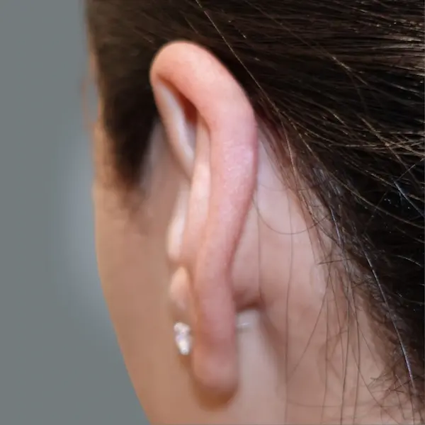
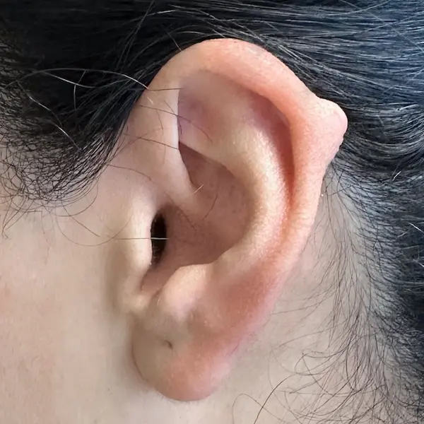
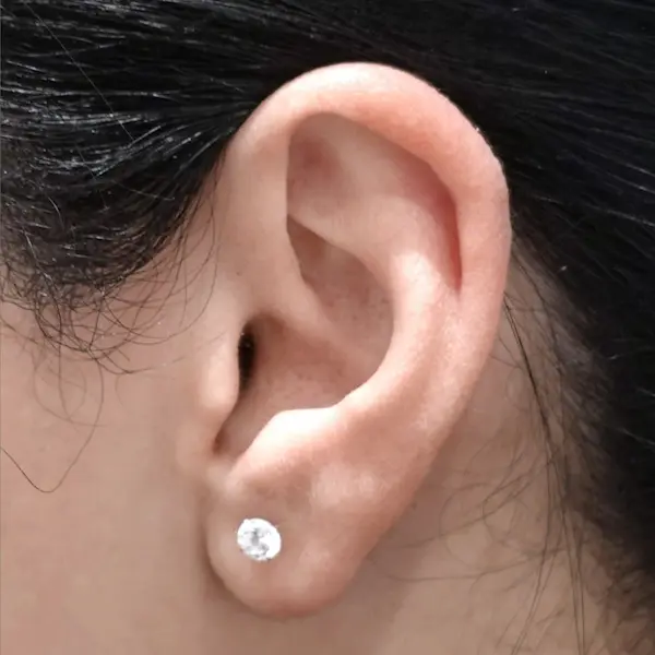
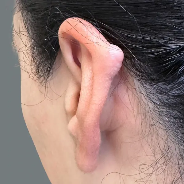
Patient No. 6 — Ear Keloid Before & After: Small Helix Keloid
This patient had a smaller keloid involving the outer rim of the ear. Both side and posterior views demonstrate the raised cartilage scar before treatment and the contour of the ear afterward.
BEFORE
AFTER
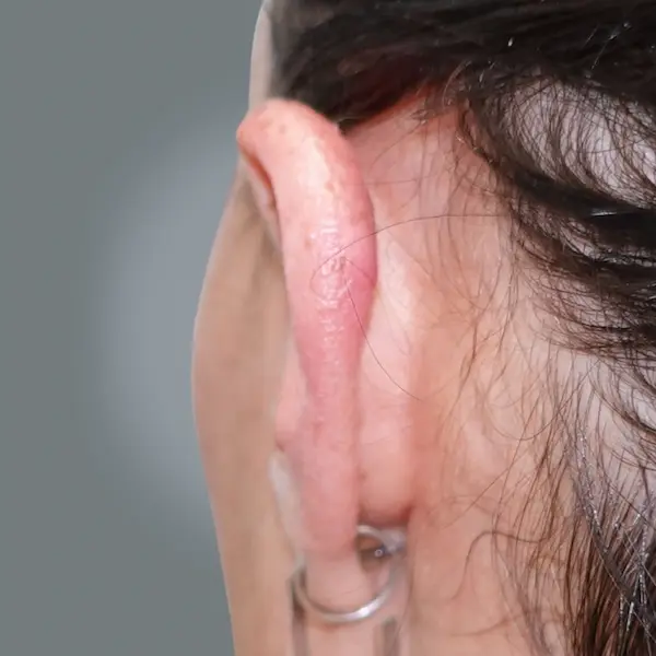
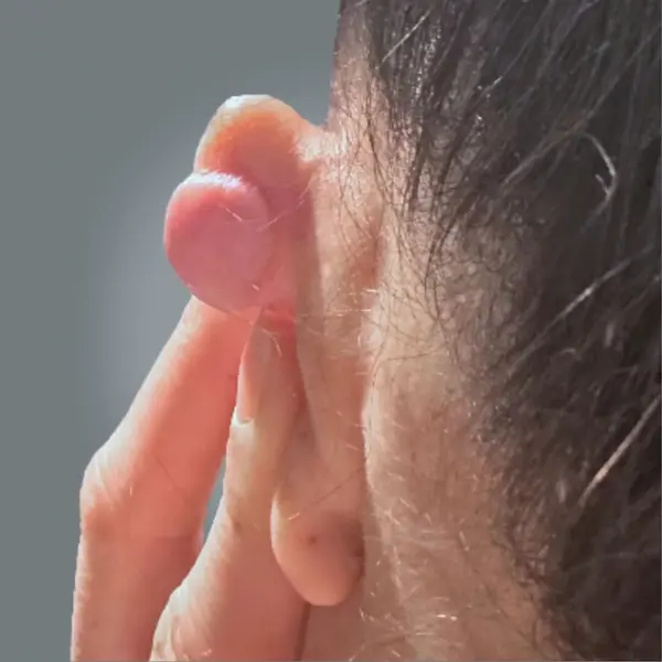
Patient No. 7 — Ear Keloid Before & After: Posterior Helix Keloid
This patient presented with a rounded keloid arising from the posterior aspect of the upper ear. The after photograph shows the treated area and restoration of the contour of the helical rim.
BEFORE
AFTER
BEFORE
AFTER
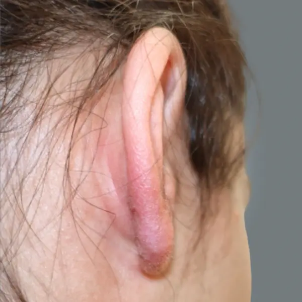
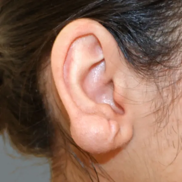
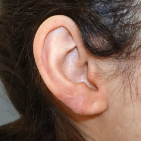
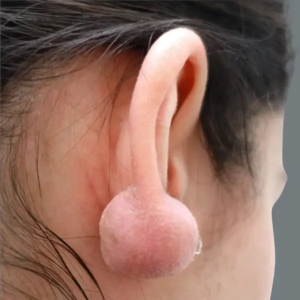
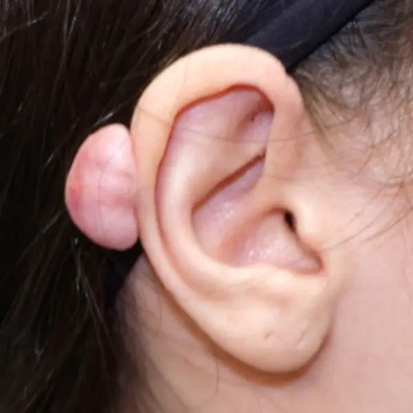
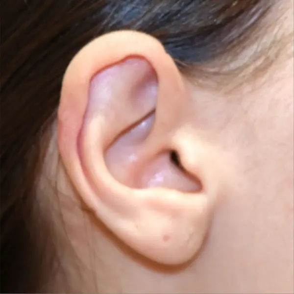
Patient No. 8 — Ear Keloid Before & After: Very Large Earlobe Keloid
This patient had a very large rounded keloid involving much of the earlobe. Because complete removal in one operation would not have left enough skin for closure, treatment was performed in stages. The side and posterior photographs show the extent of the keloid before treatment and the resulting earlobe contour after a two-stage procedure. Pressure earring and steroid treatments were followed.
BEFORE
AFTER
Patient No. 9 — Ear Keloid Before & After: Large Posterior Cartilage Keloid
This patient presented with a large keloid projecting from the posterior aspect of the upper ear. The after photograph demonstrates the upper ear contour following surgery and steroid injections.
BEFORE
AFTER
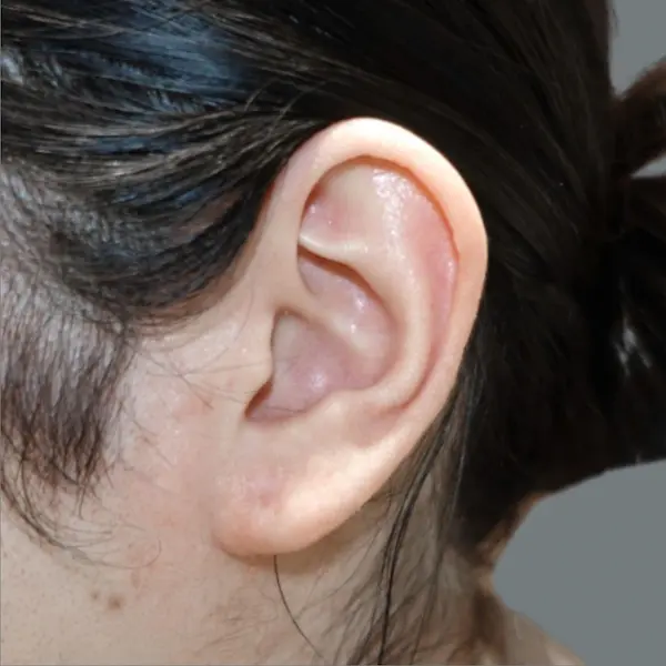
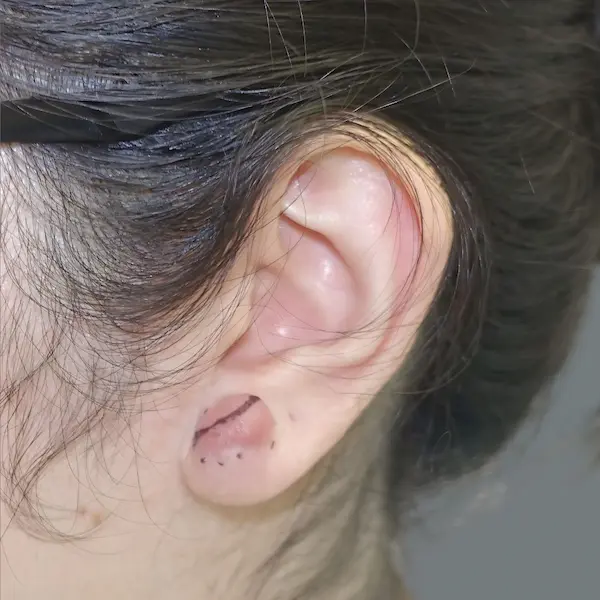
Patient No. 10 — Ear Keloid Before & After: Earlobe Keloid
This patient had a localized keloid involving the earlobe. The preoperative photograph shows the area marked for treatment, and the after image shows the earlobe after healing from surgery. Steroid treatments were continued until the scar was soft.
BEFORE
AFTER
BEFORE
AFTER
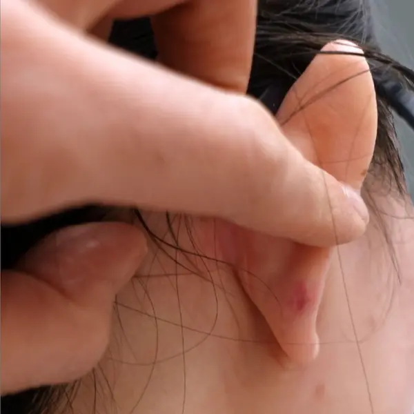
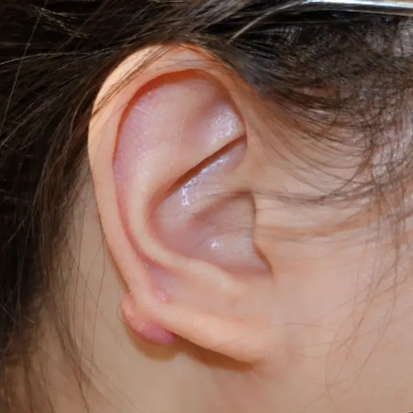
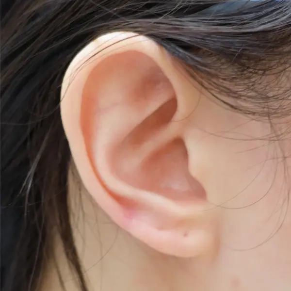
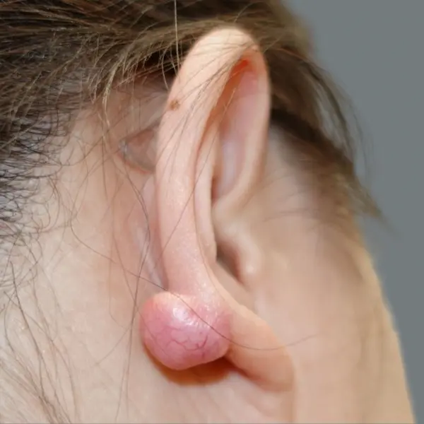
Patient No. 11 — Ear Keloid Before & After: Front and Back of the Earlobe
This patient had keloid tissue involving both the front and posterior aspect of the lower ear. Surgical removal of the posterior keloid and removal of the thickened scar on the front of the ear was performed, followed by steroid treatments.
BEFORE
AFTER
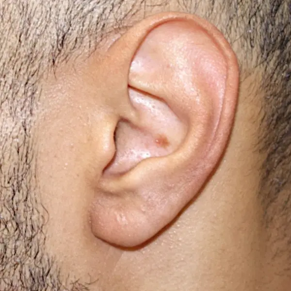
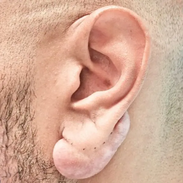
Patient No. 12 — Ear Keloid Before & After: Large Earlobe Keloid
This patient presented with a broad keloid involving a significant portion of the earlobe. The before and after photographs show the reduction of the bulky scar and preservation of the overall earlobe contour following intralesional excision and skin flap preservation to line the back of the ear, steroid treatments, and pressure earring therapy.
BEFORE
AFTER
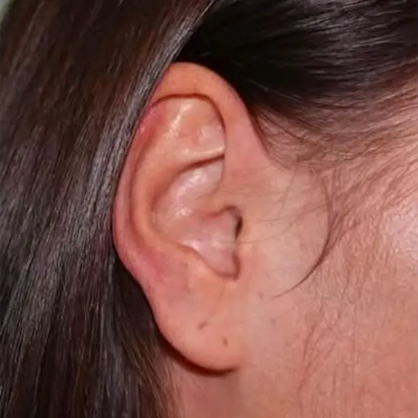
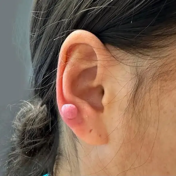
Patient No. 13 — Ear Keloid Before & After: Small Lower Ear Keloid
This patient had a smaller rounded keloid involving the lower outer portion of the ear. The after photograph shows the treated area with restoration of the surrounding ear contour.
BEFORE
AFTER
BEFORE
AFTER
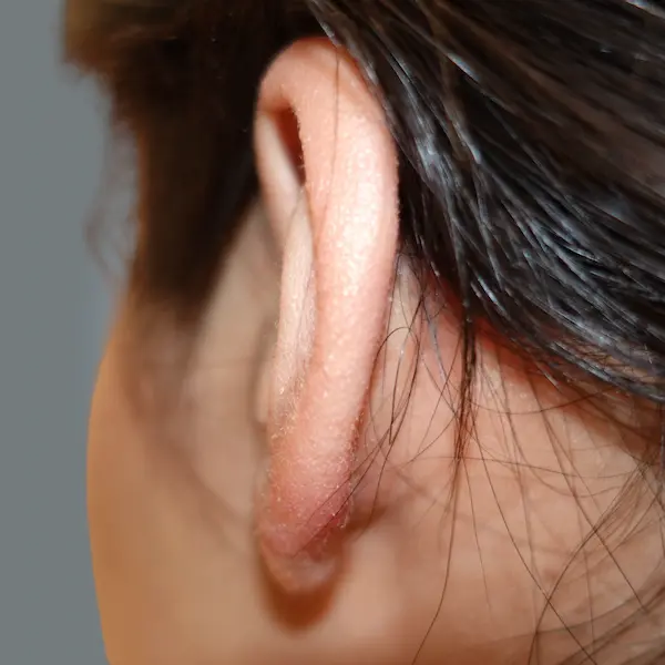
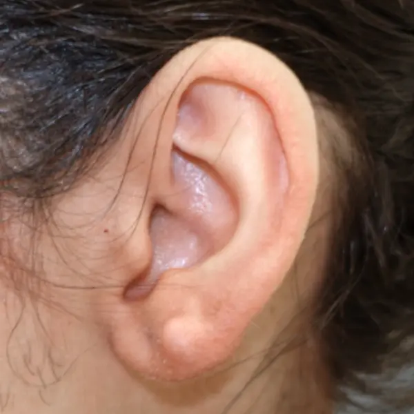
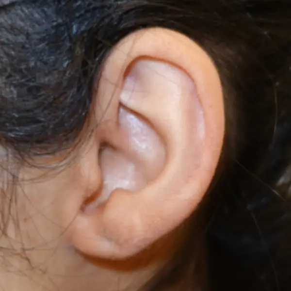
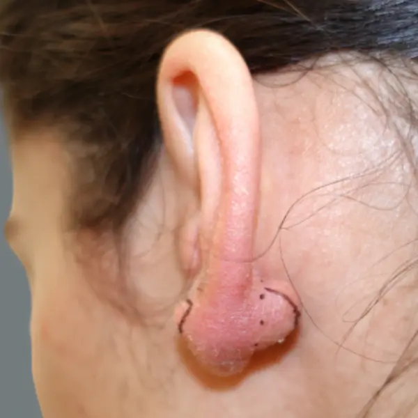
Patient No. 14 — Ear Keloid Before & After: Lower Ear Keloid, Side and Posterior Views
This patient has a keloid that extended from the front to the back of the lower ear.
An intralesional excision was performed, removing the firm central keloid while preserving skin for closure. The patient then underwent steroid injections/pressure therapy during follow-up.
